Figure 3.
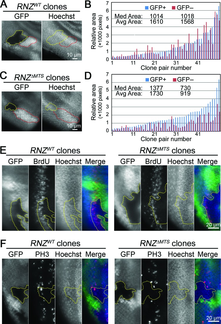
Mitochondrial dRNaseZ is autonomously required for imaginal disc cell proliferation. (A,C) Fluorescent images of third instar eye discs containing RNZWT and RNZΔMTS clones in a wild-type background. Yellow dots outline RNZWT and RNZΔMTS clones (GFP negative); red dotes outline the wild-type twin-spots (double GFP). Nuclei are stained with Hoechst. (B,D) Areas of clones and corresponding twin-spots measured in pixels with histogram function of Adobe Photoshop. Bars are ordered on the X-axis according to the size of the twin-spot (blue, GFP+). Values for median and average clone areas are indicated. RNZWT clones (B) have similar sizes with wild-type twin-spots (P = 0.67), while RNZΔMTS clones (D) are smaller (P < 0.0001, t-test). (E,F) BrdU incorporation (E) and PH3 staining (F) posterior of the morphogenetic furrow in third instar eye discs. In merge: GFP, green; BrdU or PH3, red; Hoechst, blue. GFP negative RNZWT and RNZΔMTS clones are outlined with yellow dots, and discs are oriented with anterior to the right. Note that RNZΔMTS clones have thinner BrdU band and no PH3 staining.

