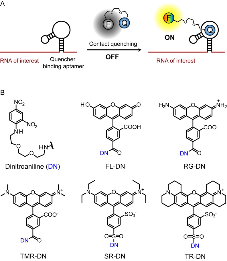
Figure 1.
Scheme for imaging RNA by using contact-quenched fluorogenic probes. (A) The contact-quenched fluorophore-dinitroaniline conjugates (OFF) light up upon binding to the quencher binding aptamer (ON). RNA of interest can be fused to the quencher binding aptamer and imaged in the presence of the fluorophore-quencher conjugate. F denotes any fluorophore and Q denotes a contact quencher. (B) Structures of the contact quencher and fluorogenic probes used in this study. Dinitroaniline (DN) is the contact-quencher used in this work. FL-DN: fluorescein-dinitroaniline, RG-DN: rhodamine green dinitroaniline, TMR-DN: tetramethylrhodamine-dinitroaniline, SR-DN: sulforhodamine-dinitroaniline, TR-DN: TexasRed-dinitroaniline.

