Figure 6.
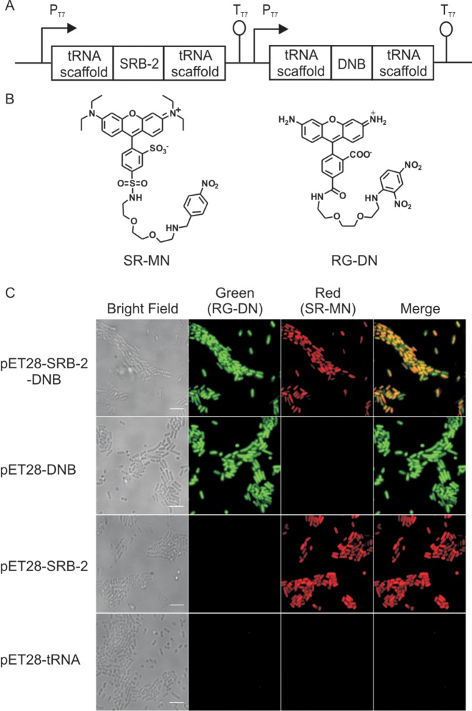
Dual colour labelling strategy using a quencher-binding aptamer and a fluorophore-binding aptamer. (A) Cloning vector used for expressing both SRB-2 and DNB (pET-SRB-2-DNB) aptamers in E. coli. (B) Structure of the two fluorogenic probes used for dual-colour imaging, SR-MN: sulforhodamine B-p-nitrobenzylamine and RG-DN: Rhodamine green-dinitroaniline. (C) Imaging DNB and SRB-2 aptamers in live E. coli with RG-DN and SR-MN (1 μM, each), respectively. Fluorescence signal in both red and green channel was detected in cells expressing both DNB and SRB-2, while cells expressing either DNB or SRB-2 showed fluorescence only in the green or red channel, respectively. Cells expressing the tRNA scaffold showed no fluorescence signal. Scale bar, 5 μm.

