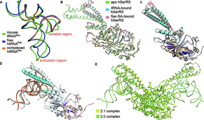
Figure 3.

Structural changes induced by tRNA binding. (A) The structural superposition of the free human tRNASec (blue, PDB 3A3A), complexed human tRNASec (orange) and the free mouse tRNASec (green, PDB 3RG5). (B) The backbone alignment of the free hSerRS (green, PDB 3VBB), Ser-SA-bound hSerRS (salmon, PDB 4L87) and tRNA-bound hSerRS in the 2:1 complex (blue). (C) The overlay of the two monomers of the 2:1 complex. There is a 6°-angle rotation in the helical TBD due to the binding of tRNA, while the ADs superimpose well. (D) The overlay of the two monomers of the 2:2 complex. One subunit is in color (tRNA in orange) as in Figure 1A while the other is in gray. (E) The backbone overlay of the ADs of the 2:2 and 2:1 complexes. hSerRS in the 2:2 complex is colored green while in the 2:1 complex is yellow.
