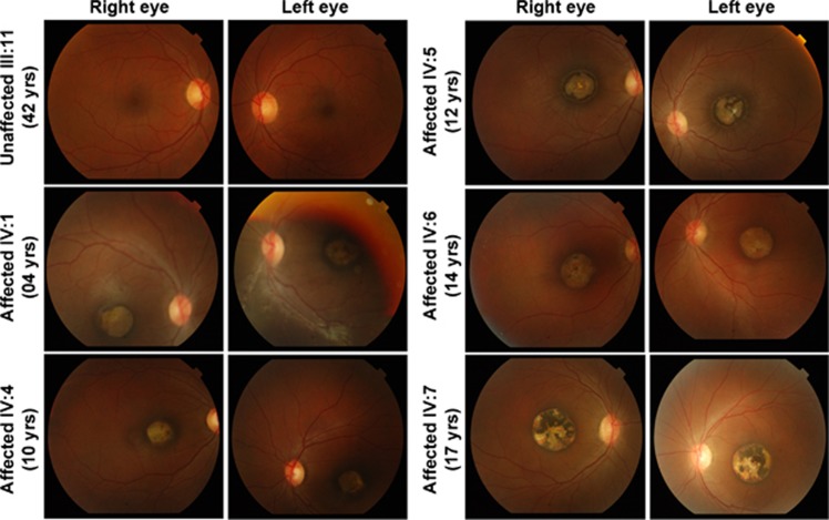
Figure 2.
Funduscopic imaging revealed macular degeneration in affected individuals from family PKAB157. Color photographs of the fundus from both eyes demonstrate macular atrophy of varying degrees in all affected individuals. This macular atrophy was centered on the fovea and included the retina, RPE and choroid, with preservation of larger, deep choroidal vessels. The areas of atrophy were surrounded by a ring of RPE hyperplasia and, in individual IV:5, radially oriented retinal striae.

