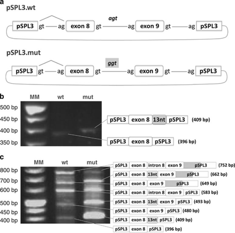
Figure 2.
Minigene constructs and transcript analysis of transfected cells. (a) schematic representation depicting the minigene constructs. The PCR fragment comprising the last portion of intron 7, exon 8, intron 8, exon 9 and the first portion of intron 9 was cloned in the pSPL3 vector: pSPL3.wt minigene presents the genomic wild-type fragment; pSPL3.mut minigene presents the genomic mutant fragment (c.820+13A>G mutation); (b) transfection of HeLa cells with the wild-type and mutant constructs; RT-PCR pattern and sequence analysis of the corresponding fragments are depicted in the schemes; (c) transfection of COS-7 cells with the wild-type and mutant constructs; RT-PCR pattern and sequence analysis of the corresponding fragments are depicted in the schemes.

