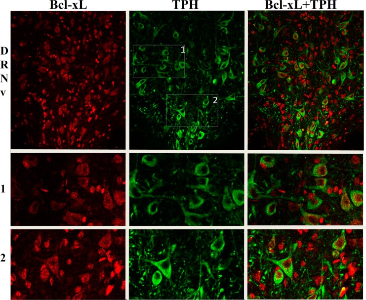
Fig 1. Representative photomicrographs of immunofluorescent staining.
Bcl-xL (red), TPH (green) and double immunofluorescence for TPH and Bcl-xL are shown in the rat ventral part of the dorsal nucleus (DRNv), and at higher magnification in the random insets 1 and 2 as indicated in the DRNv TPH picture.

