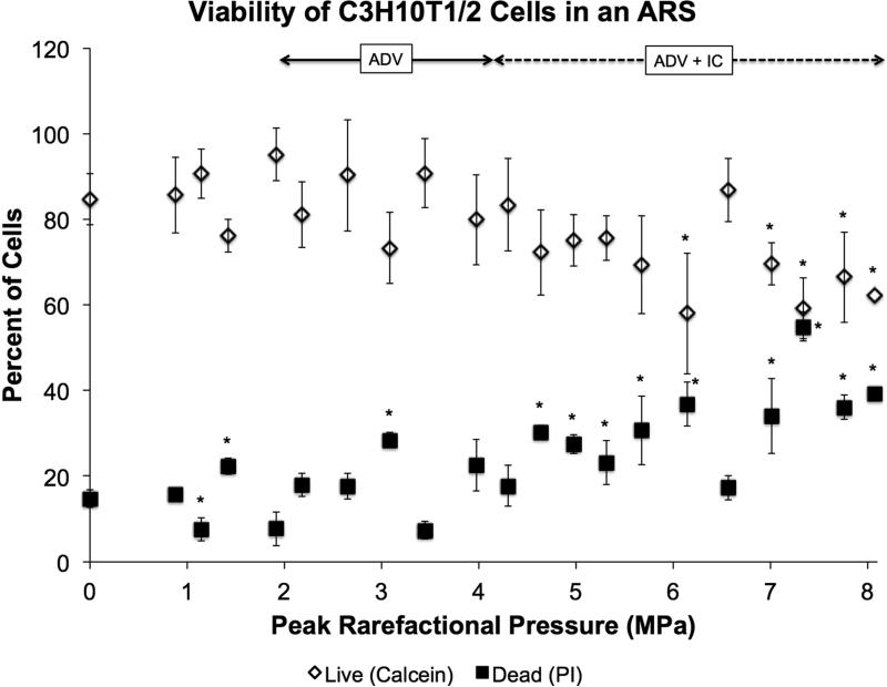
Figure 3.
Viability of C3H10T1/2 cells in an ARS containing 5 mg/mL fibrin, 1% (v/v) PFP double emulsion, and 50,000 cells/mL after exposure to US at 13 cycles and 10 Hz PRF. Viability was determined with calcein for live staining, propidium iodide for dead staining, and Hoechst for total number of cells. The sham condition (i.e., 0 MPa) underwent the same experimental steps and exposure to environmental conditions as the non-sham conditions. Data are shown as mean ± standard deviation for n = 9. *p < 0.05 vs. no US condition.

