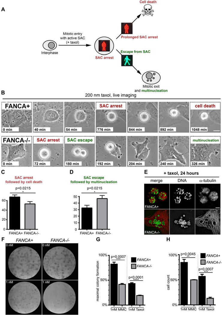
Figure 6. Loss of FANCA promotes escape from SAC and is synthetic lethal with low-dose taxol exposure.
(A) Assay schematic. Prolonged activation of SAC triggers cell death to prevent genomic instability by eliminating cells that cannot satisfy the checkpoint. Escape from SAC followed by erratic chromosome segregation and mitotic exit generates multinucleated cells. (B) Representative time-lapse imaging snapshots of FANCA+ and FANCA−/− cells exposed to taxol. Note prolonged SAC arrest followed by cell death in gene-corrected cell and escape for SAC followed by cytokinesis failure and multinucleation in FANCA−/− cell. Scale bar: 15 µm. Time from mitotic entry is shown for each frame. Time-lapse phase-contrast frames of cells grown in DMSO supplemented with 10% FBS at 37°C, 5% CO2 were acquired every 2 minutes for at least 24 hours on a Nikon Biostation live-imaging system (C, D) Quantification of time-lapse imaging experiments. FANCA−/− cells are more likely to escape SAC and less likely to be eliminated through SAC-associated death compared to gene-corrected isogenic cells (p=0.0215). Data for 115 mitotic FANCA+ cells and 129 mitotic FANCA−/− cells (three experimental replicates for each cell line) were analyzed with two-tailed t-test. See Supplemental Movies 1–2. (E) Prolonged prometaphase arrest in FANCA+ cells and multinucleation in FANCA−/− cells upon 24-hour exposure to taxol in an independent experiment. Images acquired on an Applied Precision personalDx deconvolution microscope. (F) Representative colony-forming (CFU) assay plates. Primary FANCA−/− fibroblasts and FANCA”+ fibroblasts (500 cells per 10 cm2 plate) were exposed to taxol for 11 days. Note decreased colony formation on FANCA−/− plates exposed to 1 nM of taxol. (G) Quantification of the CFU assay shown in (F). FANCA−/− cells are more sensitive to 1 nM taxol than FANCA+ cells in the CFU assay. 1 nM MMC was used as positive control. (H) Direct cell counts confirm that stable expression of FANCA rescues both taxol and MMC hypersensitivity of FANCA−/− patient cells. Two-way ANOVA with Sidak correction was used for data comparison. Data show pooled results of three separate experiments, expressed as the mean ± SEM in triplicates.

