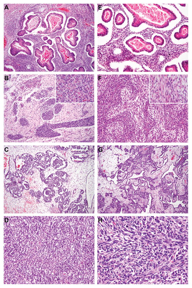
Figure 2.
DICER1 mutation status and morphology in ovarian Sertoli-Leydig cell tumors. (A–D) Representative micrographs of DICER1-mutant tumors; (E–H) representative micrographs of DICER1 wild-type tumors. (A&E) Sertoli-Leydig cell tumors with heterologous gastrointestinal-like morphology, (B&F) Sertoli-Leydig cell tumors with rhabdomyosarcomatous morphology (inset), (C&G) Sertoli-Leydig cell tumors with retiform morphology, (D&H) Poorly differentiated Sertoli-Leydig cell tumors. No association between DICER1 hotspot mutation status and ovarian Sertoli-Leydig cell tumor morphology was observed.

