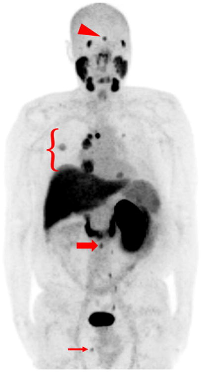
Fig. 1.

MIP image of patient #2 who was status post-right nephrec-tomy with multiple sites of metastatic disease. Normal radiotracer uptake is noted in the lacrimal glands, salivary glands, oropharynx, nasopharynx, liver, spleen, proximal small bowel, left kidney, left ureter, and bladder. Abnormal sites of uptake include a brain lesion (red arrowhead), lung nodules as well as mediastinal and hilar lymph nodes (red bracket), a retroperitoneal lymph node (thick red arrow), and a soft tissue perineal lesion (thin red arrow) (color figure online)
