
Fig. 3.
T3 vertebral body bone lesion from patient #5 which was occult on contrast-enhanced CT of the chest (a), but demonstrated moderate radiotracer uptake (SUVmax 2.6) when imaged with 18F-DCFPyL PET/CT (b) (arrowheads)
Official websites use .gov
A
.gov website belongs to an official
government organization in the United States.
Secure .gov websites use HTTPS
A lock (
) or https:// means you've safely
connected to the .gov website. Share sensitive
information only on official, secure websites.
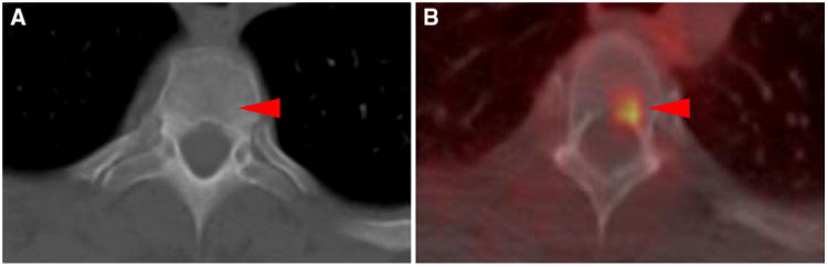
T3 vertebral body bone lesion from patient #5 which was occult on contrast-enhanced CT of the chest (a), but demonstrated moderate radiotracer uptake (SUVmax 2.6) when imaged with 18F-DCFPyL PET/CT (b) (arrowheads)