Fig. 4.

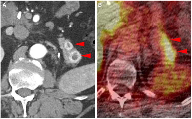
Metastatic lesions to the pancreatic tail in patient #1 (SUVmax 5.0 in the more anterior lesion and SUVmax 7.4 in the more posterior lesion) as noted on contrast-enhanced CT (a) and 18F-DCFPyL PET/CT (b) (arrowheads)

Metastatic lesions to the pancreatic tail in patient #1 (SUVmax 5.0 in the more anterior lesion and SUVmax 7.4 in the more posterior lesion) as noted on contrast-enhanced CT (a) and 18F-DCFPyL PET/CT (b) (arrowheads)