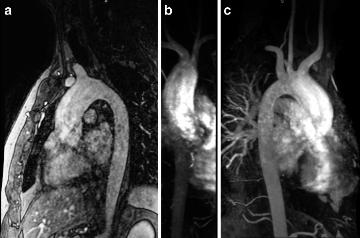
Fig. 17.
Contrast-enhanced MR angiography of the aorta in a 50-year-old man with a bicuspid aortic valve and moderate stenosis, complicated by mild insufficiency and ascending aorta dilatation. a T1-weighted 3-D spoiled gradient echo after a timed bolus contrast of gadolinium, b-c maximum intensity projection reconstructions

