
Figure 2.
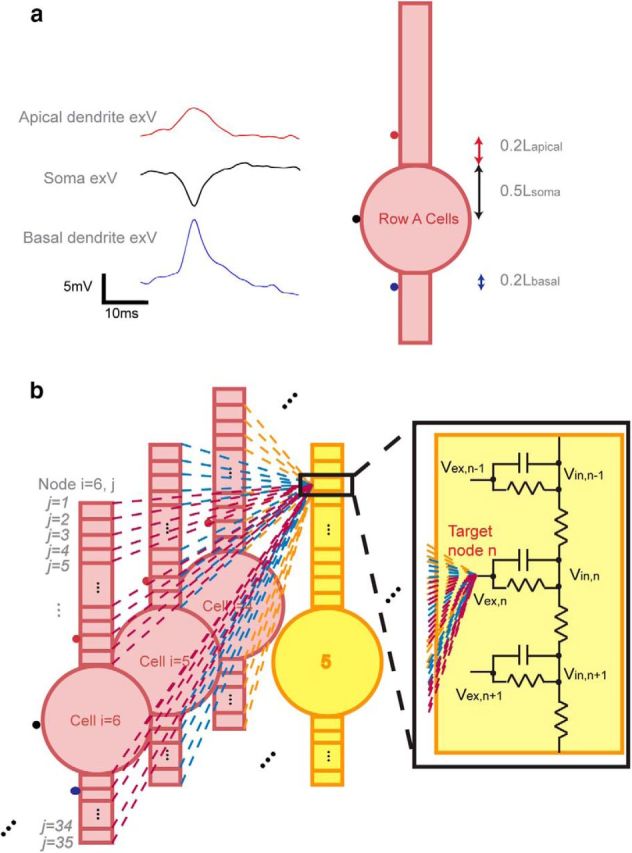
Electric field interactions within the neural network. a, Initiation of the action potential by extracellular stimulation for Row A cells using in vitro acquired signals. Each of the three signals was 10 times the average of 10 randomly selected trials from in vitro acquired signals (ie, cell layer, apical dendrites, basal dendrites). This is because physiological signals were acquired 10–30 μm away from its nearest cells due to the damage caused by the electrode insertion (estimated to be 2–3 cells layers), whereas in the model, signals were inserted directly on the cell membrane. As extracellular potential decays at a rate inversely proportional to the distance away from a point source, the potential amplitudes in the model were expected to be larger than what were seen experimentally. The signals were applied at the middle point of soma and at the dendritic compartments adjacent to the soma. b, Network field effect acting on one extracellular node of a target cell using Equation 2. The field acting on node n of cell 5 in Row B was the superposition of all fields generated by all Row A cells.