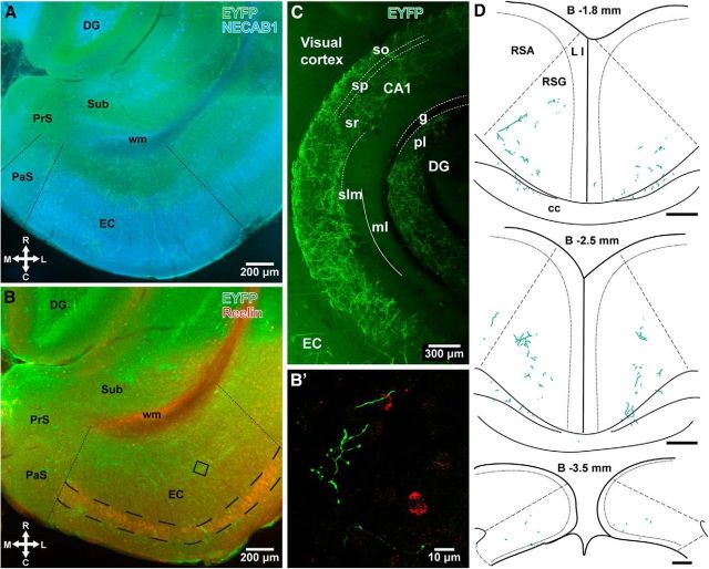
Figure 3.
Identification of the target regions of EYFP-labeled septal axons by immunohistochemistry in the hippocampus and extrahippocampal areas. A, B, Epifluorescent images showing demarcation (dashed lines) of the parasubiculum (PaS) and entorhinal cortex (EC) by immunohistochemistry for NECAB1 (blue), and the EC by reelin (red) in horizontal brain sections of a PV-Cre mouse superimposed with EYFP-immunofluorescent septal axons (green). The EC and PaS are respectively differentiated by dense and lighter NECAB1 immunoreactivity (A; case GU41s47). Immunolabeling for reelin visualizes mEC LII (large dashed lines) by labeling the stellate cells and interneurons (B; case GU41s20). The white matter (wm) is labeled nonspecifically. B shows, in a confocal microscopic image (maximum intensity projection, 5.14 m z-stack), anterogradely labeled septal axons (green) near reelin-positive interneurons (red, from the framed area in B). C, Differential innervation of layers of the hippocampus by EYFP-labeled medial septal axons; maximum intensity confocal microscopic projection of a 350-μm-thick z-stack of a Clarity-processed thick section. Solid line marks the hippocampal fissure between the CA1 region and dentate gyrus (case GU31hpc). D, Anterogradely labeled septal PV-expressing GABAergic axons at three rostrocaudal levels in the retrosplenial cortex (animal GU42). Dashed lines indicate estimated borders between the agranular (RSA) and granular (RSG) retrosplenial cortices. Scale bars, 200 μm. Median filter was applied (x, y: radius, 1 pixel) in C. DG, dentate gyrus; PrS, presubiculum; Sub, subiculum; L I, layer I; so, stratum oriens; sp, stratum pyramidale; sr, stratum radiatum; slm, stratum lacunosum-moleculare; ml, molecular layer of the dentate gyrus; g, granule cell layer; pl, polymorphic layer of the dentate gyrus; cc, corpus callosum; R, rostral; C, caudal; M, medial; L, lateral; B, bregma.

