Figure 10.

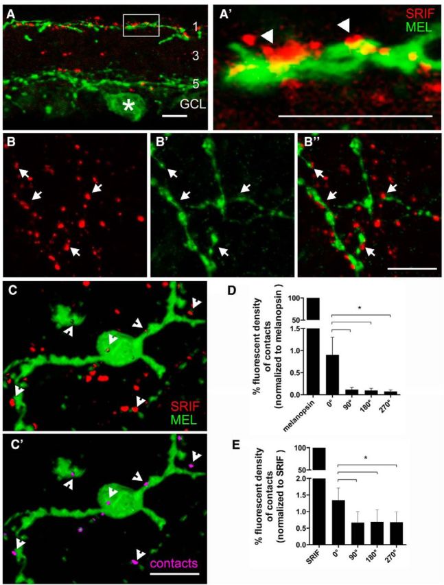
SRIF and melanopsin immunostaining show the juxtaposition of SRIF amacrine cell processes and M1 ipRGC dendrites in stratum 1 of the IPL. A, SRIF amacrine cell processes (red) are mainly in stratum 1 of the IPL and a few processes are in strata 3 and 5 of the IPL. A melanopsin-immunoreactive somata (asterisk) and its dendrites in strata 1 and 5 of the IPL. A′ is a magnification of the inset in A showing melanopsin ipRGC dendrites and SRIF amacrine cell processes forming contacts in stratum 1 of the IPL. B, SRIF-immunoreactive varicose processes in stratum 1 of the IPL in a whole mount. B′, Melanopsin immunoreactivity shows robust labeling of melanopsin ipRGC varicose dendrites. B″, Merged images show close contacts between SRIF amacrine cell processes often near the varicosities of the M1 ipRGC dendrites. C, A 3D reconstruction of SRIF processes and melanopsin dendrites from an image stack taken through the IPL of a retinal whole mount shows many punctate SRIF amacrine cell processes near a displaced melanopsin-immunoreactive ipRGC somata and dendrites. C′, Omitting the red channel that labeled SRIF amacrine cells reveals the contacts (pink) formed with melanopsin ipRGC somata and dendrites. Arrows show the contacts between SRIF amacrine cell processes and melanopsin ipRGC dendrites. D, Quantification of fluorescent density created from the contacting processes was normalized to the fluorescent density of the melanopsin immunoreactivity. Rotated images had significantly less fluorescent density generated from the contacting processes. E, Fluorescent density of the contacts was also normalized to the fluorescent density of SRIF immunoreactivity. Fluorescent density of contacts after rotation was significantly less than un-rotated image. Scale bars, 10 μm. *p < 0.05.
