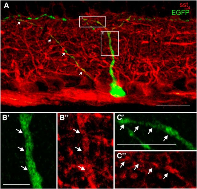
Figure 11.
Transverse section through an OPN4–EGFP retina showing colocalization of EGFP fluorescence with sst4 immunoreactivity. A, OPN4–EGFP retinas contain EGFP fluorescent (green) M1 ipRGCs. sst4 immunoreactivity (red) is localized to RGC somata and to numerous processes throughout the IPL and RGC axons in the NFL. Inset B is magnified in B′ and B″ showing an M1 ipRGC dendrite ascending to stratum 1 of the IPL that is colabeled with sst4 immunoreactivity. Inset C is magnified in C′ and C″, which shows colocalization between M1 ipRGC and sst4 immunoreactivity in stratum 1 of the IPL. Arrows indicate the colocalized OPN–EGFP fluorescent process with sst4 immunoreactivity. Scale bars: A, 20 μm; B, 5 μm; C, 10 μm.

