Figure 5.

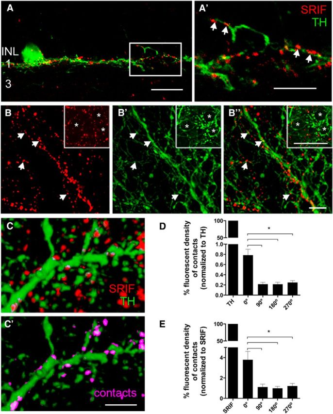
SRIF and TH immunostaining show closely apposed SRIF- and TH-immunoreactive amacrine cell processes in stratum 1 of the IPL. A, SRIF amacrine cell processes (red) and TH-immunoreactive DA amacrine cell processes (green) are in close proximity in stratum 1 of the IPL. A′, A 3× magnification of the box in A reveals multiple contacts between SRIF amacrine cell and DA amacrine cell processes in stratum 1 of the IPL. Whole-mount retina shows varicose and fine SRIF amacrine cell processes (B) and large- and fine-caliber DA amacrine cell processes (B′). B″, Merged images highlight the juxtaposition of the SRIF amacrine and DA amacrine cell processes. Contacts are more frequent near the varicosities of the DA amacrine cell processes. Arrows show the points of contacts between SRIF and DA amacrine cell processes. Insets show perisomatic rings formed by SRIF and TH immunoreactivity. Asterisks mark the cell bodies that are surrounded by SRIF and DA amacrine cell processes. C, A 3D model of SRIF and DA amacrine cells from an image stack taken through the IPL of a retinal whole mount was generated to determine contacts between the processes. C′, Omitting the red channel reveals SRIF-immunoreactive processes and puncta that make contact with DA amacrine cell processes. Pink labeling shows contacts between SRIF and DA amacrine cell processes. D, Quantification of fluorescent density created from the contacting processes was normalized to the fluorescent density of the TH immunoreactivity. Rotated reconstructions (90°, 180°, and 270°) had significantly less fluorescent density generated from the contacting processes. E, Fluorescent density of the contacts was also normalized to the fluorescent density of SRIF immunoreactivity. Fluorescent density of contacts after rotation was significantly less than the unrotated image. Scale bars: A, 20 μm; A′, B, C, 10 μm. *p < 0.05.
