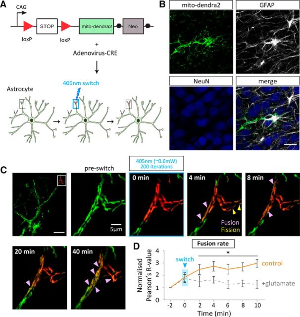
Figure 4.
Glutamate decreases the fusion rate of mitochondria in astrocytic processes in situ. A, Schematic representation of CRE-driven mito-dendra2 expression in hippocampal slices. A mouse line expressing floxed-stop-mito-dendra2 infected with an AV-CRE allows astrocyte-specific expression. B, Expression is shown to be astrocyte-specific as mito-dendra-2 colocalizes with GFAP (shown in merge). Scale bar, 20 μm. C, Representative image of mito-dendra2 expression with 18 μm2 region of activation, before and 4–40 min after 200 iterations of 20% 405 nm laser light (∼0.6 mW). ROI shows example fusion dynamics under basal conditions in a distal astrocytic process. Scale bar, 20 μm; ROI, 5 μm. D, Quantification of the rate of fusion under basal conditions (control) and in the presence of glutamate (100 μm + 1 μm glycine). Single focal-plane Pearson's R values (au) represent colocalization of green and red mito-dendra2 signal before and after switching with 405 nm laser light and subsequently imaged every 2 min (this is defined as the fusion rate, ie, change in R value over a 10 min period; control: n = 5 cells, 5 slices; glutamate: n = 5 cells, 5 slices). *p < 0.05.

