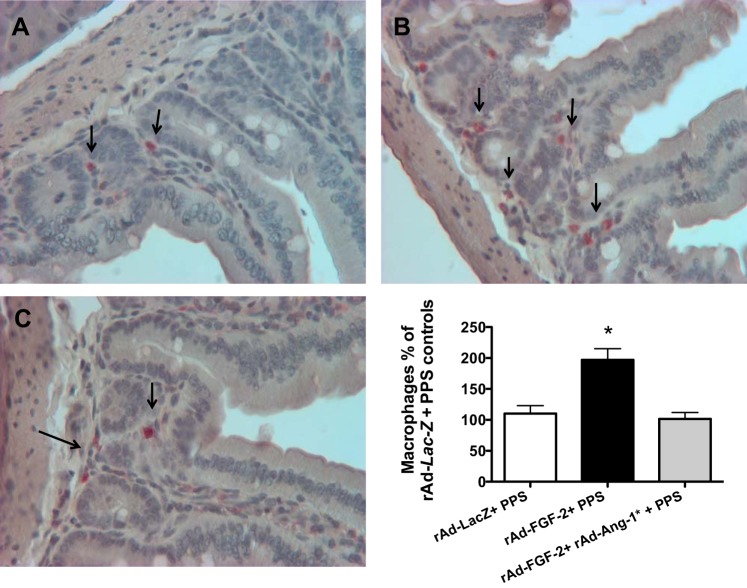
Fig. 8.
Ang-1* decreases the intestinal recruitment of macrophages induced by FGF-2 + PPS. A–C: a representative immunohistochemistry staining of macrophages in control mice treated with either rAd-Lac-Z +PPS (A); mice treated with rAd-FGF-2 + PPS (B); and mice treated with rAd-Ang-1* + rAd-FGF-2 + PPS (C). Macrophages are shown in red color stained with a rat anti-mouse antibody against the F4/80 antigen. All sections were counterstained with hematoxylin. The graph represents the quantification of macrophages expressed as % changes relative to control mice treated with rAd-Lac-Z + PPS. *Kruskal-Wallis test, P < 0.0082, n = 4 per group; Dunn's Multiple Comparison Test: rAd-Lac-Z + PPS vs. rAd-Ang-1* + rAd-FGF-2 + PPS, P > 0.05; and rAd-Lac-Z + PPS vs. rAd-FGF-2 PPS, P < 0.05. Original magnification (A–C), 200×.

