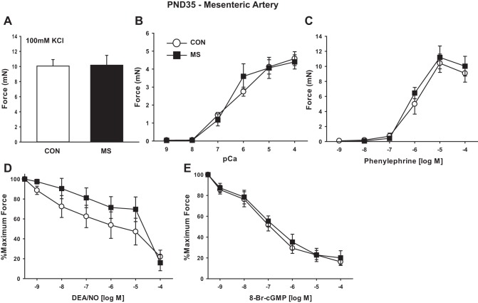
Fig. 7.
MS stress does not alter contractile function of MAs at PND35. Rats were stressed and MA1s studied as described above. Data are plotted as developed force in mN (A–C), or as percentage of force remaining in response to cumulative doses of vasodilator in arteries preconstricted at submaximal concentrations of PE (10 μM) (D–E). A: force generated to maximal KCl-induced depolarization (100 mM). B: dose response to calcium after α-toxin permeabilization. C: dose response to the α-adrenergic agonist PE. D: dose response to NO donor DEA/NO in intact arteries. E: dose response to 8-Br-cGMP in α-toxin permeabilized MA1s activated at submaximal calcium (pCa6). Values are means ± SE with n = 5–6/group.

