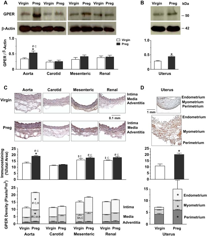
Fig. 4.
Protein amount and tissue distribution of GPER in the aorta, carotid artery, mesenteric artery, and renal artery as well as the uterus of virgin versus pregnant rats. Tissue homogenates of blood vessels (A) and the uterus (B) were prepared for Western blots using GPER antibody (1:1,000). GPER and ERα (shown in Fig. 2) were run on the same gel and therefore have the same actin control. Vascular tissue sections (C) and uterine sections (D) were stained with GPER antibody using immunohistochemistry, and the total amount and relative distribution of GPER brown immunostaining in different layers of the tissue wall were measured using ImageJ. Bar graphs represent means ± SE; n = 4–6 rats/group. *P < 0.05, preg vs. virgin rats; ‡significantly different (P < 0.05) from corresponding measurements in the aorta, carotid artery, mesenteric artery, and renal artery of virgin rats; #significantly different (P < 0.05) from corresponding measurements in the aorta, carotid artery, mesenteric artery, and renal artery of pregnant rats.

