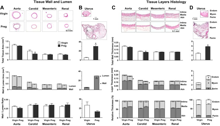
Fig. 5.
Histology and morphometric analysis of the aorta, carotid artery, mesenteric artery, and renal artery (A and C) as well as the uterus (B and D) of virgin versus pregnant rats. Cryosections were prepared for hematoxylin and eosin staining, and tissue images were analyzed using ImageJ. Total tissue area, lumen area, whole wall area, and wall-to-lumen ratio were measured. Total wall thickness and the relative thickness of the different vascular layers (intima, media, and adventitia) and uterine layers (endometrium, myometrium, and perimetrium) were also measured. Bar graphs represent means ± SE; n = 4–6 rats/group. *P < 0.05, pregnant vs. virgin rats.

