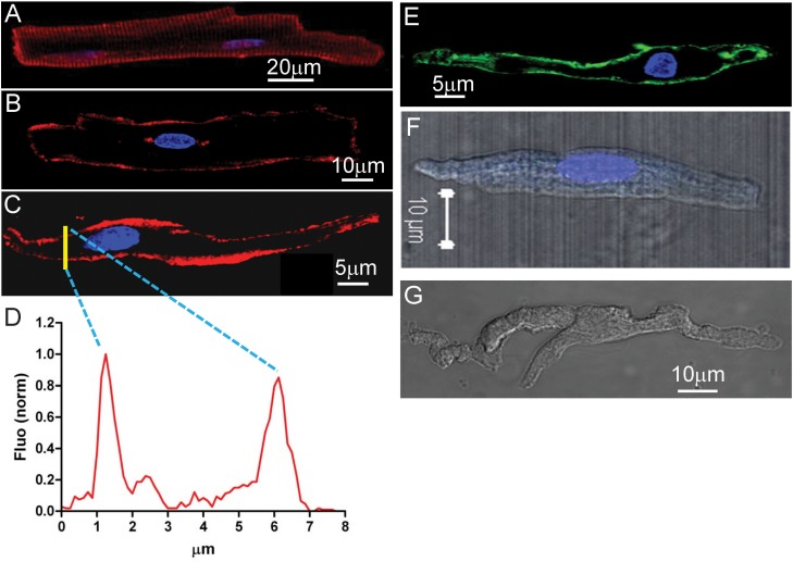
Fig. 2.
Leptin receptor expression at surface of cardiac myocytes isolated from in SD rat. A: a ventricular myocyte (transverse scanning image focused on cell surface). B: an atrial myocyte (transverse scanning image focused on the nucleus). C: a sinus node myocyte (transverse scanning image focused on the nucleus). Leptin receptor is shown in red, and the nucleus is shown in blue (DAPI). D: yellow line in C marks a section of transverse scanning image to reveal the distribution of fluorescence shown in the graph. Two peaks indicate the plasma membrane expression of leptin receptors. E: HCN4 expression at cell surface of a sinus node myocyte. F: lack of HCN4 expression without use of HCN4 antibody in an atrial myocyte as a negative control. G: lack of leptin receptor expression without use of leptin receptor antibody in sinus node myocytes as a negative control.

