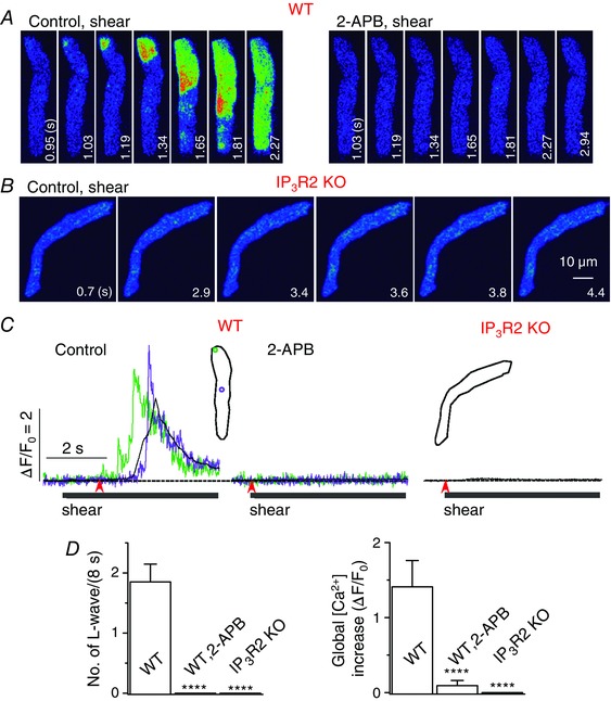
Figure 2. IP3R2 is responsible for triggering longitudinal Ca2+ waves under shear stress .

A and B, confocal Ca2+ images recorded in the representative WT (with and without 2‐APB) (A) and IP3R2 KO mouse atrial myocytes (B) during the application of shear stress (∼16 dyn cm−2). Longitudinal Ca2+ waves were not observed during shear application when IP3R2 was deficient. C, Ca2+ fluorescence measured from the ROIs with corresponding colours (inset) from the series of confocal images recorded in the cells illustrated in A and B. The time marked by arrowheads matches with 0 s shown in the confocal images. D, summary of the number of longitudinal Ca2+ waves and whole‐cell Ca2+ increases during 8‐s application of shear stress in WT cells (n = 15) with and without 2‐APB, and in IP3R2 KO cells (n = 12). ****P < 0.0001 vs. WT (unpaired t test).
