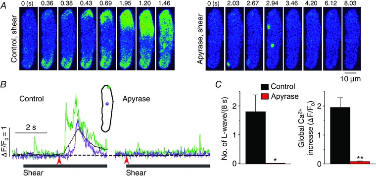
Figure 7. ATP released to extracellular space elicits longitudinal Ca2+ waves during shear stress .

A, representative confocal Ca2+ images recorded during the applications of shear (∼16 dyn cm−2) in the absence (‘Control, shear’) and presence of the ATP metabolizing enzyme, apyrase (2 U ml‒1; ‘Apyrase, shear’). This enzyme did suppress the occurrence of longitudinal Ca2+ waves under shear stress. B, changes in local (green and violet) and global (black) Ca2+ levels measured from the ROIs (inset) on the series of confocal Ca2+ images recorded from the cell shown in A. The time marked by arrowheads matches with 0 s shown in the confocal images. C, summary of the effects of apyrase (n = 8) on the occurrence of longitudinal Ca2+ waves and on global Ca2+ changes during the application of shear (8 s). *P < 0.05, **P < 0.01 vs. Control (paired Student's t test).
