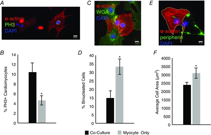
Figure 3. Sympathetic innervation of cardiomyocytes in culture inhibits cardiomyocyte maturation .

A, representative image showing a proliferating cardiomyocyte double‐labelled with α‐actinin and phospho‐histone H3 (PH3; scale bar = 10 μm). B, bar plot showing a higher rate of proliferation for cardiomyoctes co‐cultured with sympathetic neurons for 5 days (black) than for cardiomyocytes grown in isolation (grey; n = 4). C, representative image showing a binucleated cardiomyocyte identified by α‐actinin, staining of cell membranes with wheat germ agglutinin (WGA), and DAPI nuclear staining (scale bar = 10 μm). D, bar plot showing co‐cultures (black) had a decreased number of binucleated cardiomyocytes compared to myocyte‐only cultures (grey; n = 3). E, representative image showing a cardiomyocyte, visualized with α‐actinin staining, outlined with ImageJ, neurons visualized with peripherin, and DAPI nuclear staining (scale bar = 10 μm). F, cardiomyocytes co‐cultured with sympathetic neurons for 4 days (black) were smaller than cardiomyocytes that were cultured for the same period in isolation (grey; n = 3; *P < 0.05 vs. co‐culture.)
