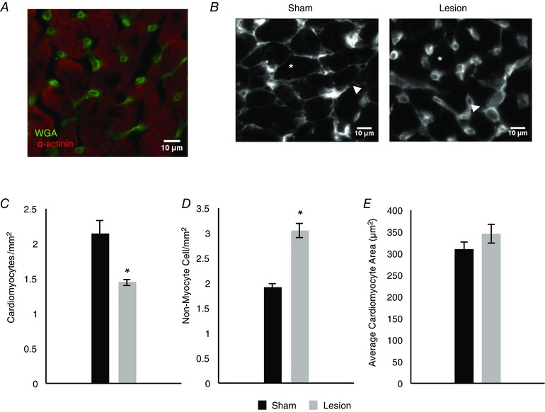
Figure 9. Sympathetic innervation is required for the regulation of cardiomyocyte density in vivo .

A, representative image of wheat germ agglutinin (WGA) and α‐actinin staining in ventricular sections of 8‐week‐old rats. WGA delineates the cellular membrane of cardiomyocytes as well as staining non‐myocyte cells in the slice. B, representative images of WGA staining showing cardiomyocytes (asterisk) and non‐cardiomyocyte cells (arrowhead) in sham (left) and lesioned (right) animals. C, bar plot showing that, compared to sham (black), neonatal sympathetic lesions (grey) result in a decrease in cardiomyocyte density in 8‐week‐old animals (n = 3). D, bar plot comparing the density of non‐myocyte cells in sham (black) and lesioned (grey) animals, showing an increase in the density of non‐myocyte cells in lesioned animals (n = 4). E, bar plot showing that the average cardiomyocyte size in sham (black) and lesioned (grey) animals is not significantly different, suggesting that the difference in heart size in lesioned animals cannot be accounted for by decreased cell size. (n = 3; *P < 0.05 vs. sham.)
