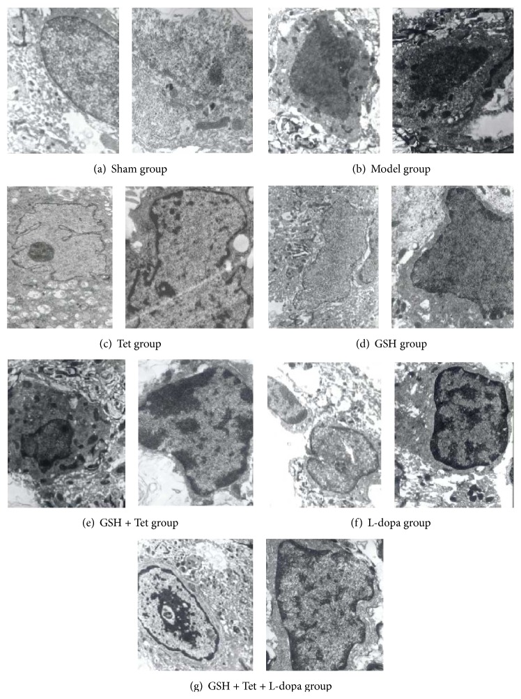
Figure 6.
Transmission electron microscopy (TEM) images of apoptotic dopaminergic neuron in the substantia nigra pars compacta (SNc) of lesion sides. Images for all groups were taken using the same magnification (in one group, the left at 11,000x, the right at 15,000x). Dopaminergic neurons with normal cellular morphology were detected in the sham group. The model group showed characteristics of apoptosis in the early PD stage. The L-dopa group, however, displayed characteristics of apoptosis in the middle-late PD stage. In the Tet group and GSH groups, changes of apoptosis were less obvious compared with the L-dopa group. In the GSH + Tet and GSH + Tet + L-dopa group, several dopaminergic neurons were in preapoptosis stages.

