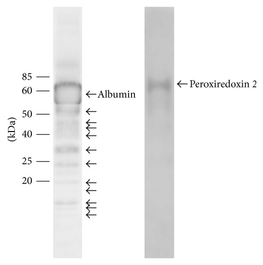
Figure 3.

Western blot analysis of sham rabbit vitreous developed with anti-albumin and anti-peroxiredoxin 2. Labeled arrows correspond to the respective full length proteins. The unlabeled arrows for the lower molecular mass bands on the anti-albumin blot may indicate the specific cleavage fragments, which correspond with some of the differentially expressed 2D-PAGE protein spots.
