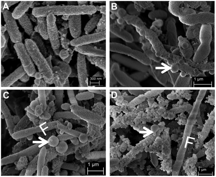
FIGURE 2.
Representative scanning electron micrographs (SEM) of SMK279a cells grown in the presence or absence of ampicillin at 37°C for 48 h on LB agar plates. SEM images of SMK279a cells were recorded as previously published (Krohn-Molt et al., 2013). (A) SEM image of cells obtained from colonies cultured in the absence of ampicillin. (B) SEM image of cells obtained from small colonies cultured in the presence of 100 μg/ml ampicillin. (C) SEM image of cells obtained from big colonies cultured in the presence of 100 μg/ml ampicillin. (D) SEM image of cells obtained from big colonies cultured in the presence of 300 μg/ml ampicillin. Cells from (A–C) predominantly formed long filamentous cells (indicated by letter F) and OMVs (indicated by arrows). In the presence of ampicillin, SMK279a cells became enlarged and formed OMVs with sizes up to 677 nm in diameter.

