FIGURE 4.
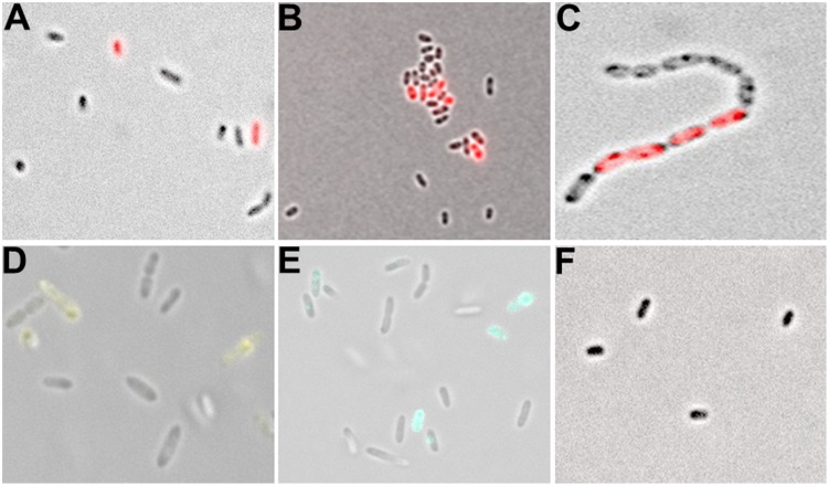
Analysis of single cell fluorescence of PblaL1 and PblaL2 promoter gene fusions. (A) Expression of the blaL1 promoter fused to rfp in SMK279a. Cells were grown at 30°C for 17 h under aerobic conditions (200 rpm) in LB medium containing 100 μg/ml ampicillin. Thereby, 2.0 ± 0.72% cells were in the bla-ON and 98.0 ± 0.72% cells were in the bla-OFF mode. (B) Expression of the blaL2 promoter fused to rfp in SMK279a grown under the same conditions as indicated in (A). Here, 4.4 ± 0.69% of cells were in the bla-ON and 95.6 ± 0.69% cells were in the bla-OFF mode. (C) Phenotypic heterogeneity observed in cells (carrying the PblaL2::rfp fusion) forming long cell chains. Cells were grown overnight under aerobic conditions (120 rpm) in LB medium supplemented with 100 μg/ml ampicillin. (D) Phenotypic heterogeneity observed in cells carrying the PblaL2::yfp (E) and the PblaL2::cfp promoter gene fusion. (F) Control cells of SMK279a were grown under the same condition as described in (A) carrying a promoterless rfp reporter fusion (Pless::rfp). Images were recorded with 63x/1.30 Oil M27 and 100x/1.30 Oil M27 lenses using a Zeiss Axio Imager 2 fluorescence microscope (Zeiss, Jena, Germany) and employing appropriate filters.

