. 2015 Nov 13;7(4):505–508. doi: 10.4055/cios.2015.7.4.505

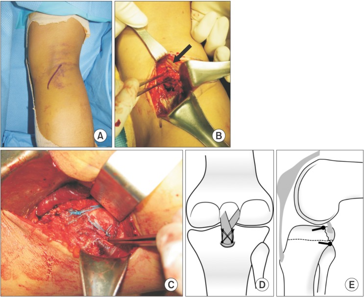
Fig. 1. Posterior view of the right knee. (A) An oblique incision between semitendinosus tendon and medial head of the gastrocnemius. (B) The medial head of the gastrocnemius (arrow) is held laterally to expose the posterior capsule of the knee joint. Subsequently, capsulotomy with a longitudinal L-shaped incision is made and extended laterally to expose the fracture site. The forceps hold a bony fragment of the posterior cruciate ligament tibial attachment. (C) Fixation technique of the suture bridge method, with proximal to the left and distal to the right of the photo. (D) Illustration showing the position of the anchors in the coronal plane. (E) Illustration showing the position of the anchors in the sagittal plane.

Fig. 1