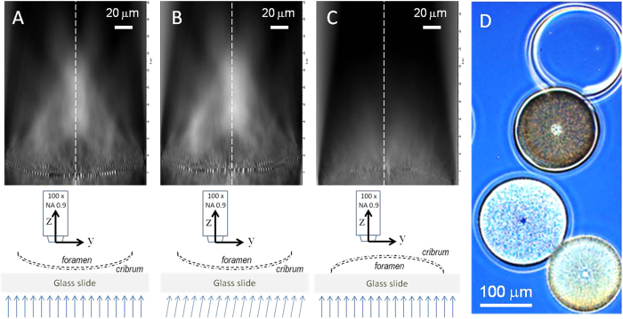
Figure 3. Hyper-maps of a single valve of Coscinodiscus centralis under different orientations.
The cribrum faces a normal incident light (A). The cribrum faces a 10° tilted incident light (B). The foramen faces a normal incident light (C). Light microscopy picture of cleaned valves, where the effect of orientation is visible as light valve (like in A, cribrum side facing the incoming light (like in A,B) and a dark valve (like in (C), foramen facing the incoming light) (D). In each map, the spectral envelope is normalized to the light source intensity. The schemes describe the measurement configurations. The vertical dotted lines indicate the symmetry axis of the valve (S). The scale bar (20 μm) is the same for the Figure (A–C).

