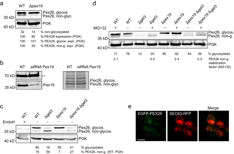
Figure 5. The Pex19-dependent steps occur after or independently of ER entry of PEX26.
(a,b) ER integration of PEX26-ops is independent of PEX19, both, in HeLa and in yeast cells. (a) Knock-out of PEX19 does not affect the ER entry of PEX26-ops but affects the level of non-glycosylated PEX26. Expression of PEX26-ops in Δpex19 cells for 120 min. To compare protein expression in wild-type and Δpex19 PEX26-ops expression is stated in relation to PGK levels. ‘%PEX26 expression’ is the total amount of PEX26 relative to PGK. ‘%PEX26, glycos. expr.’ and ‘%PEX26 non-g. expr.’ are the levels of glycosylated and non-glycosylated forms relative to PGK. WT was set to 100%. Quantifications show that only the non-glycosylated PEX26-ops is reduced to 35% in Δpex19 cells. (b) PEX19 does not influence ER entry of PEX26-ops in mammalian cells. HeLa cells expressing PEX26-ops were treated for 60 hours with siRNA directed against PEX19 or with non-targeting siRNA. For radiolabelling, cells were grown for 15 min with [35S]-cysteine and [35S]-methionine before immunoprecipitation of PEX26-ops. Western blot (left): Knockdown of PEX19. The asterisk marks a nonspecific band as a loading control. Autoradiograph of radio-pulse experiment (right): Pex26-ops glycosylation is not reduced. (c) Pex19 stabilizes cytosolic PEX26-ops in the absence of Get3. The additional Δpex19 knockout in Δget3 reduces the non-glycosylated form of PEX26-ops from 59% to 27%. ‘%PEX26 non-g. (WT, PGK)’ is relative to wild-type, normalized by PGK protein expression. Both parts of each panel were cropped from the same blot. (d) Pex19 and Get3 stabilize cytosolic PEX26-ops. Non-stabilized PEX26-ops is degraded by the proteasome. Cells treated with 30 μM MG132 show a 2.1- to 4-fold increase of non-glycosylated, cytosolic PEX26-ops in wild-type, Δpex19, Δget3, and Δpex19Δget3 strains. (e) Pex19 and Get3 are required for organelle targeting of PEX26. Localization of EGFP-PEX26 in Δpex19Δget3 yeast. GAL1 promoter-driven expression was recorded 80 min after chase. EGFP-PEX26 does not co-localize with the ER marker Sec63-RFP. Bar = 5 μm. Full-size blots and autoradiograph are presented in Supplementary Figure S1.

