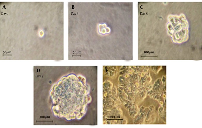
Figure 1. Generation Spheroid Cells From HT-29 Cell Line.
(A-D): Representative photographs have been showing colon cancer cells expanded in vitro as undifferentiated spheres in serum-free medium containing EGF and FGF-2. Pictures have shown a typical colon sphere observed by the inverted phase contrast microscope. (e): Microscopically analysis of colon cancer spheres have cultivated in differentiation conditions (growth factors removal and addition of 10% FBS) for 7 days (200 ×).

