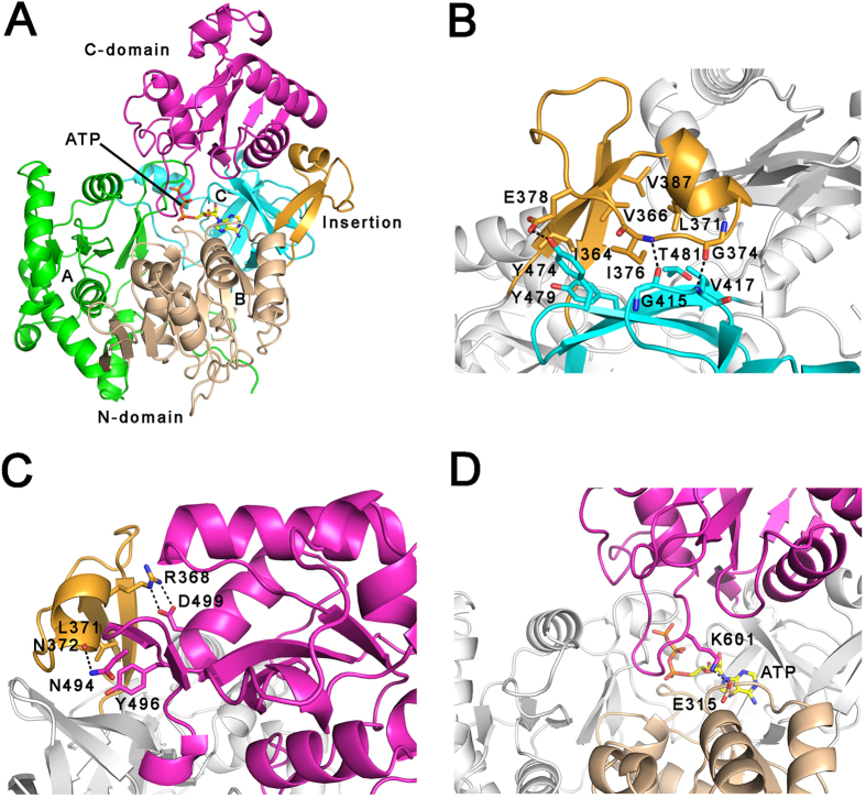
Figure 1. Overall structure of ATP-bound MsFadD32.
(A) Ribbon representation of the MsFadD32 structure. The three subdomains of the N-terminal domain are colored green, wheat, and cyan, respectively. The insertion motif and C-terminal domain are colored orange and magenta, respectively. ATP is shown as yellow sticks. (B) The interactions between the insertion motif and N-terminal subdomain C. (C) The interactions between the insertion motif and C-terminal domain. (D) The ATP-bound MsFadD32 adopts an atypical adenylate-forming conformation. In (C–D), the residues involved are shown as sticks and the hydrogen bonds are shown as dashed lines.

