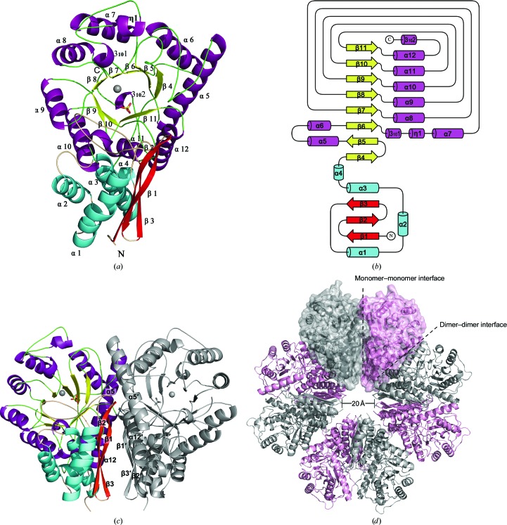
Figure 2.
Overall structure of Sa_enolase. (a) Ribbon diagram of the overall structure of Sa_enolase. The secondary-structural elements are coloured cyan/red for the N-terminal domain and purple/yellow for the C-terminal barrel domain. The α-helices and β-strands are labelled in black. (b) Topology diagram of Sa_enolase. The secondary-structural elements are indicated. (c) The dimeric structure of Sa_enolase. Secondary-structure elements (β1–β3, α5 and α12) involved in dimerization are labelled in black. (d) The octameric structure of Sa_enolase. The monomers within a dimer are shown in grey and pink. The monomer–monomer interface and dimer–dimer interface are highlighted with black dashes.

