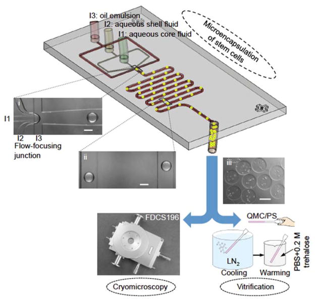
Figure 1.
A schematic illustration of the experimental setup and procedure. The procedure includes three major steps: microencapsulation of murine embryonic stem cells (mESCs) or human adipose-derived stem cells (hADSCs) using a nonplanar microfluidic flow-focusing device, cryomicroscopy study of inhibiting devitrification and intracellular ice formation (IIF) using a Linkam cryostage, and vitrification study of cell survival and function with quartz microcapillary (QMC) and conventional plastic straw (PS). The insets (i), (ii), and (iii) are typical images of microdroplets at the proximal of the flow-focusing junction, alginate hydrogel microcapsules in the serpentine channel, and collected stem cell-laden microcapsules, respectively. The microcapsules are highly monodisperse (~220 μm) as the droplet pinching-off falls into the dripping mode of flow instability. LN2: liquid nitrogen. PBS: phosphate buffered saline (isotonic by default). Scale bars: 200 μm.

