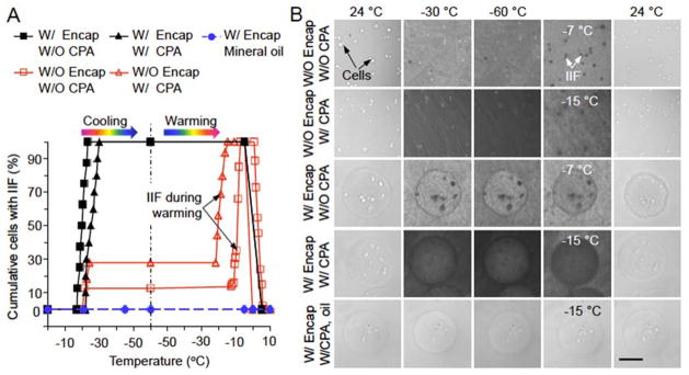
Figure 2.
Intracellular ice formation (IIF) in mESCs from cryomicroscopy studies. (A) The cumulative percentage of cells with IIF under five different conditions: (1), without (W/O) encapsulation (Encap) and W/O cryoprotectant (CPA), (2), W/O Encap and with (W/) CPA, (3), W/Encap and W/O CPA, (4), W/Encap and W/CPA in culture medium, and (5), W/Encap and W/CPA in mineral oil. The CPA is a combination of 1.5 M 1,2-propanediol (PROH, penetrating) and 0.5 M trehalose (non-penetrating). Both the cooling and warming rates are 60 °C min−1. (B) Typical images of mESCs during cooling and warming under the same five conditions in (A). The cells with and without IIF can be identified by the dark and bright spots, respectively. Scale bar: 100 μm.

