Fig. 1.

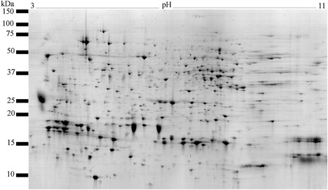
A representative gel image of the male accessory gland proteome of C. maculatus. The proteome was separated by 2D IEF SDS-PAGE and stained with Colloidal Coomassie blue

A representative gel image of the male accessory gland proteome of C. maculatus. The proteome was separated by 2D IEF SDS-PAGE and stained with Colloidal Coomassie blue