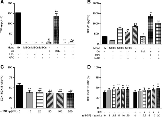
Fig. 2.

Decreased TNF-α and increased TGF-β1 in hepatocytes co-cultured with MSCs related to apoptosis. a TNF-α secretion of mono-/co-cultured hepatocytes and NPc-/HPc-MSCs. b TGF-β1 secretion of mono-/co-cultured hepatocytes and NPc-/HPc-MSCs. c Neutralisation with anti-TNF inhibits cellular apoptosis and, to a lesser extent, total death of mono-cultured hepatocytes, switching the main cell death mode from apoptosis to necrosis. d Neutralisation of MSCs with anti-TGF-β1 induces cellular apoptosis and, to a lesser extent, total death of co-cultured hepatocytes, switching the main cell death mode from necrosis to apoptosis. Values are mean ± standard deviation (n = 6). *p < 0.05 and **p <0.01, versus control mono- or co-culture; ^^p <0.01, versus co-culture with HPc-MSCs; ## p <0.01, versus control mono-culture; ++ p <0.01, versus co-culture. α-TGF-β1 Anti-transforming growth factor-β1, α-TNF Anti-tumour necrosis factor, CCK18 Caspase-cleaved cytokeratin 18, CK18 Cytokeratin 18, Co Co-culture, HPc Hypoxia-preconditioned, Hx Hepatocyte mono-culture, ind. Indirect, Mono mono-culture, MSC Mesenchymal stem cell, NAC N-acetylcysteine, NPc Normoxia-preconditioned, UD undetectable
