
Fig. 3.
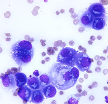
Cytology of pleural fluid. Clumps of neoplastic cells with anisocytosis, anisokaryosis, binucleations, single to multiple prominent and variably shaped nucleoli, and cytoplasmic vacuoli. The presence of a thick brush border and the “mesothelial slits” suggest a mesothelial origin of cells. May-Grünwald Giemsa staining, Obj ×60