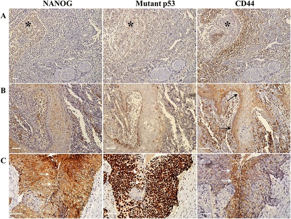
Fig. 2.

Immunostaining of well (a & b) and poorly (c) differentiated OSCCs. a In serial sections, the negative expression of NANOG, mutant p53, and CD44 is detected in one specimen of well-differentiated OSCC (* indicates cancer tissue). b In another case of well-differentiated OSCC, NANOG and mutant p53 are almost negative, seen in less than 25 % of positive cells, whereas CD44 is weakly detected in the cancer cell membrane (arrows). c In a poorly differentiated OSCC specimen, enhanced expression of NANOG, mutant p53, and CD44 is detected. The arrows indicate co-localization of the three marker proteins. Scale bar = 50 μm
