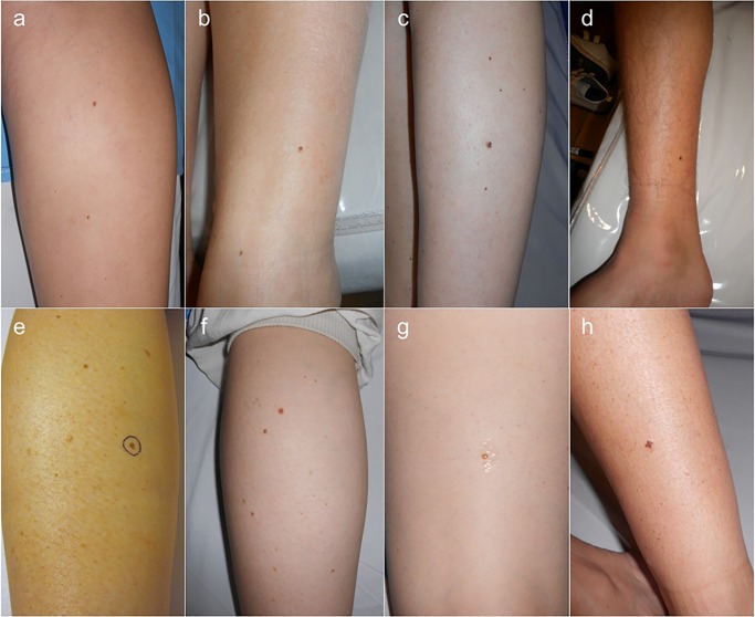
Figure 1.
Clinical images of cases 1 to 8 (a to h). Cases 1 to 4 (a to d) corresponded to melanomas detected during routine nevi control while cases 5 to 8 (e to h) corresponded to melanomas detected during digital follow-up; cases 6 and 7 (f and g) corresponded to the same patient. None of the lesions fulfilled clinical criteria for suspicion of melanoma. [Copyright: ©2015 Salerni et al.]

