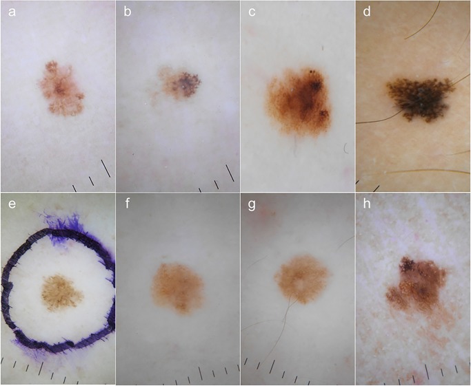
Figure 2.
Dermoscopic features of cases 1 to 8 (a to h). Reticular pattern was seen in all cases but in case 5 (e). In cases 1 to 4 (a to d) and in case 8 (h) the presence of atypical pigment network was observed. Cases 1, 4 and 5 (a, d and e) showed pseudopods at the periphery. Cases 6 and 7 displayed typical pigment network with hypo pigmented irregular areas. [Copyright: ©2015 Salerni et al.]

