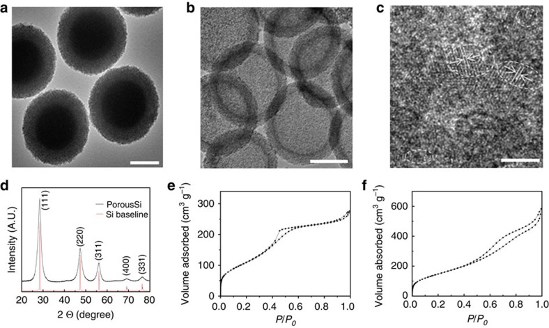
Figure 2. Structure characterizaion of the hp-SiNSs.
(a) A TEM image of solid core/mesoporous shell SiO2 particles. (b) A low magnification TEM image and (c) a high-resolution TEM image of hp-SiNSs, showing the Si particles are mostly amorphous, with scattered nanocrystalline domains with a (111) interplanar spacing of 3.13 Å. (d) X-ray diffraction patterns of the hp-SiNSs. (e,f) N2 isotherms for solid core/mesoporous shell SiO2 particles and the hp-SiNSs, respectively. Scale bar, 200 nm (a,b) 4 nm (c).

