
Figure 2.
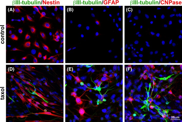
The expression of neural differentiation markers induced by taxol in C6 cells as demonstrated by immunofluorescent staining. Control (untreated) C6 cells and 100 nmol/L taxol‐treated C6 cells were analyzed by immunofluorescent staining at 48 h. In control cells, (A) the neural stem cell marker nestin was detected, (B) very few cells carrying GFAP protein could be found, and (C) immunoreactivites for β III‐tubulin and CNPase were difficult to detect. (D–F) The immunoreactivities for neuronal marker β III‐tubulin, astrocyte marker GFAP, and oligodendrocyte marker CNP, were clearly identified in the C6 cells after treated with taxol. These results indicate that expression of the neural differentiation markers were induced when the cells were challenged by taxol. Bar scale = 25 μm.