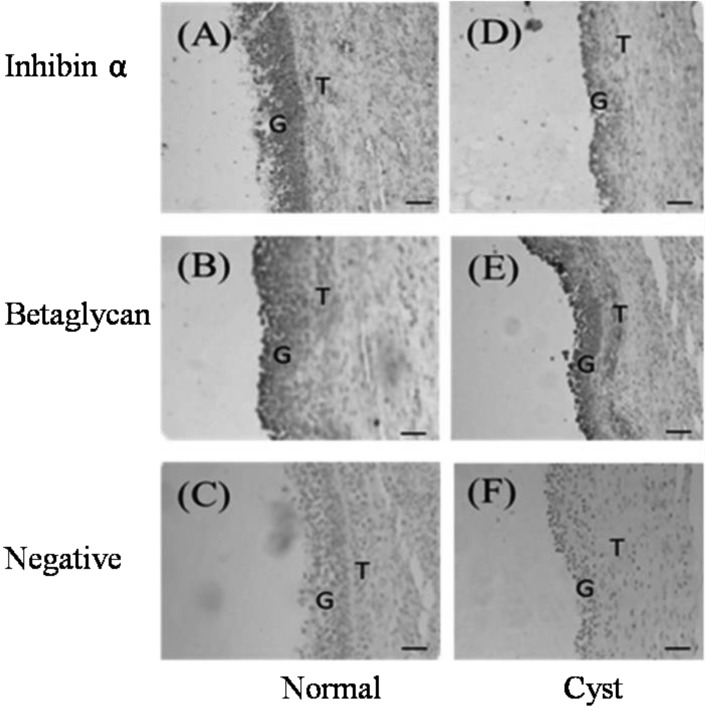
Fig. 1.
Immunohistochemical localization of inhibin α and betaglycan in porcine follicles (original magnification × 100). Brown indicates the presence of the specified protein. (A–C) Normal large follicles: anti-inhibin α IgG (A), anti-betaglycan IgG (B) and negative control (C). (D–F) Cystic follicles: anti-inhibin α IgG (D), anti-betaglycan IgG (E) and negative control (F). G indicates granulosa cells, and T indicates theca cells.

